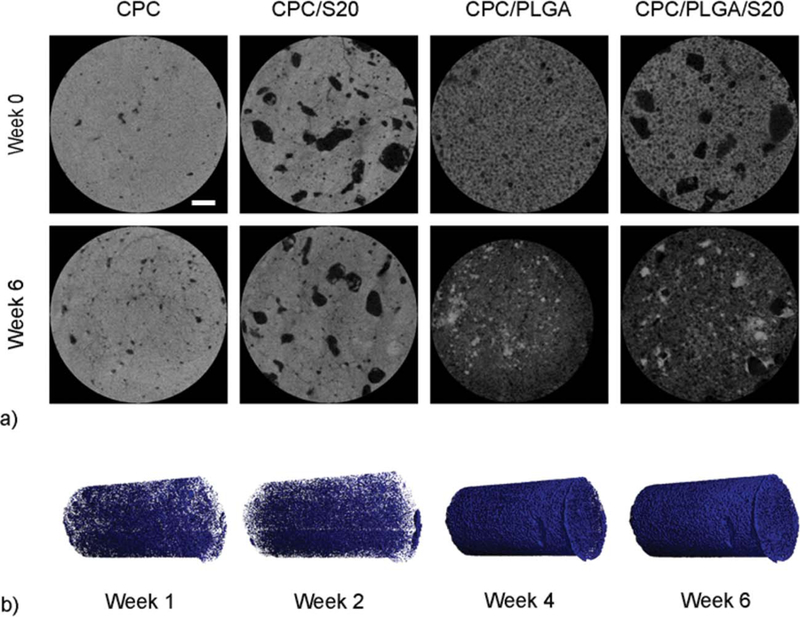
FIGURE 5.

(a) Representative microCT images of CPC, CPC/S20, CPC/PLGA, and CPC/PLGA/S20 formulations before incubation (week 0) and after 6 weeks incubation (white line represents 0.5 mm). (b) Three-dimensional reconstructed microCT of CPC/PLGA/S20, in which blue voxels represent pore formation within one sample over time.
