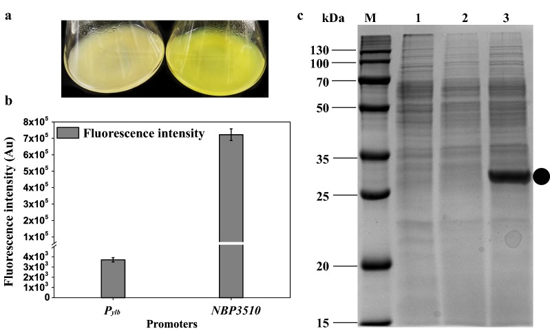
Fig. 4.
Intracellular expression of sfGFP. a The fluorescence imagines of strains WBGFP (Pylb) and WBSGFP (NBP3510). Strains were cultured for 16 h in LB medium and imagines were taken. b The fluorescence intensity controlled by different promoters. c The accumulative sfGFP protein in different strains. M, Marker. Lane 1, Strain WB800. Lane 2, Strain WBGFP. Lane 3, Strain WBSGFP. Equal amounts (30 μg) of total protein were loaded into each lane. The band corresponding to sfGFP was marked. All cultures were grown in triplicate, and each experiment was performed at least twice. Error bars indicate standard deviations

