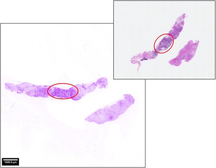
Fig. 2.
Human breast biopsy imaged with the HistologTM Scanner (left) and the corresponding H&E microscopy slide used for pathological final assessment (right). Staining with a fluorescence dye was performed before on-site scanning. The images were displayed with an artificial coloring of the grey values, which mimics an H&E stain and is adapted to the needs of the clinical users. The encircled areas indicate invasive breast cancer location as detected by the pathologists in both images using the full resolution zooming feature to reveal morphological details

