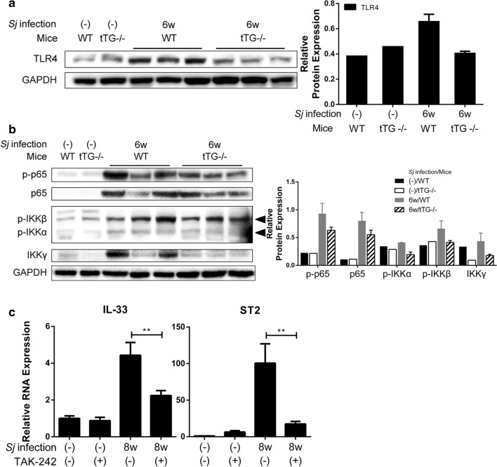
Fig. 6.
TLR4 and NF–κB pathway activation are involved in tTG-regulated IL-33/ST2 expression. a The protein expression level of TLR4 in indicated mice liver lysates detected by Western blotting is shown on the left panel, and the result of semi-quantitative analysis is displayed on the right panel. b The protein expression levels of p-p65, p-65, p-IKKα/β and IKKγ detected by Western blotting are shown on the left panel, and the result of semi-quantitative analysis is presented on the right panel. c The relative RNA expression levels of IL-33 and ST2 in liver tissue of mice infected with Sj for 6 weeks with or without exposure to the TLR4 inhibitor TAK242 were measured by RTqPCR. Gapdh was used as an internal reference [t-test, **P = 0.0069 (right panel); **P = 0.0057 (left panel)]

