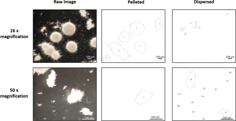
Fig. 2.
Exemplar automated image analysis of fungal macromorphologies from submerged cultures. 1 x 10⁶ spores/mL of aplD conditional expression mutants were grown in MM for 72 h at 30 °C with 220 RPM. Raw images were captured at both 16× and 50× magnification, and subsections of whole images are shown. Scale bars in lower right corners depict 500 µm. For each raw image, two quality control images are generated, in which fungal structures are depicted as outlines indexed with a unique number (red), enabling simple assessment of automated calls by the end user. One outlined image contains pellets and the other dispersed mycelia objects. Note that pelleted or clumped morphologies partially captured at the image edge are excluded from analysis. Processed outlines of fungal structures passing default definitions of pelleted (≥ 500 µm2) and dispersed (< 500 µm2 and ≥ 95 µm2) are shown for 16× magnification. Alternatively, for 50× magnification, pellet sizes definitions were identical, but dispersed mycelium were defined as < 500 µm2 and ≥ 20 µm2. We found that decreasing the lower size limit (i.e., from ≥ 95 to ≥ 20 µm2) enabled accurate automated calls for the dispersed hyphal fragments depicted at higher magnification

