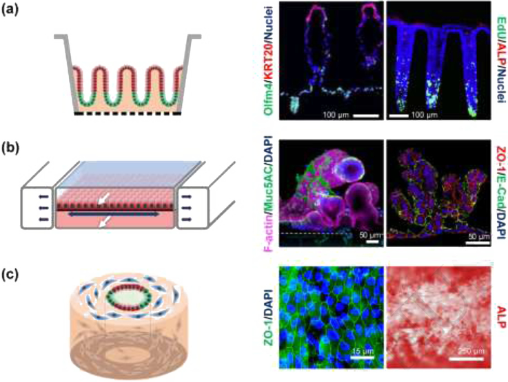
Figure 4. Shaped three-dimensional intestinal systems.
(a) Microstructured systems. Left: Side-view schematic of polarized intestinal crypts with a stem cell niche (green) and differentiated cell zone (red). Middle: Fluorescence image of cross section through an in vitro human small intestine epithelium showing a crypt and two villi. Reproduced with permission from [76]. Right: Fluorescence image of cross section through an in vitro human colon epithelium with three crypts. Stem/proliferative cells are marked by olfactomedin-4 (OLFM4) and differentiated cells by cytokeratin-20 (KRT20). Reproduced from [18] under a Creative Commons license. (b) Microfluidic systems. Left: Side-view schematic of differentiated epithelial cells (red) on a stretchable surface. White arrows mark fluid flow while dark arrows indicate the motion of the stretchable surface. Middle and right: Human small intestinal epithelial cells derived from the organoids of biopsied intestinal tissues (middle), reproduced from [77] under a Creative Commons license, and iPSC derived intestinal epithelial cells (right), reproduced from [78] under a Creative Commons license. Both tissues were grown in microfluidic devices with luminal and basal fluid flow leading to villi formation. (c) Macrostructured replica. Left: Schematic of an angled view of a silk scaffolding with fibroblasts (blue), proliferative epithelial cells (green) and differentiated epithelial cells (red). Middle and right: Fluorescence image of tight junction (ZO-1) staining and alkaline phosphatase (ALP) for human small intestinal cells grown in tubular silk scaffold embedded with myofibroblast. Reproduced from [80] under a Creative Commons license.

