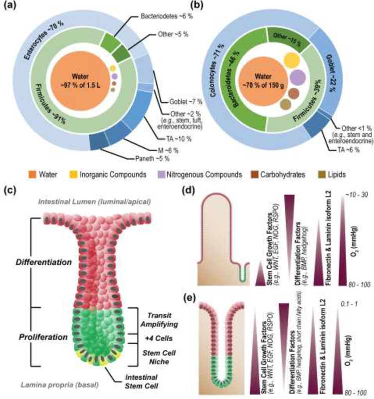
Figure I. Comparison of human small and large intestine.
Luminal contents and epithelial cell types of the small (a) and large (b) intestine [22, 101–105]. Structure of the crypts of the small and large intestines, with major zones and stem cell niche components labelled (c) Chemical gradients across the epithelium of the small and large (d) and large (e) intestine [106]. Images reproduced with permission from the indicated references.

