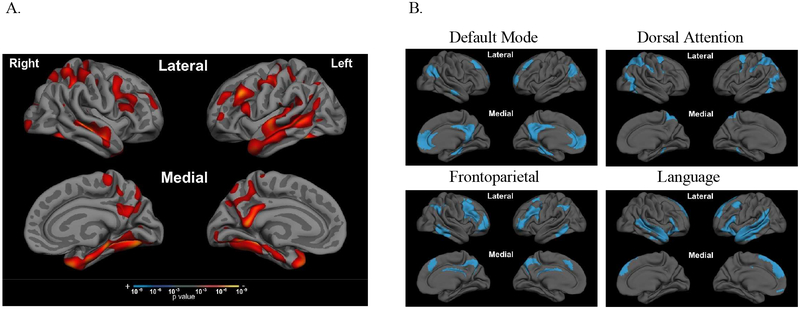
Figure 1. aMCI+ patients have cortical atrophy in multiple large-scale cortical networks outside of the MTL systems.
Compared to age-matched biomarker-negative controls (CN-), whole-brain cortical thickness analysis of aMCI+ (A) revealed atrophy in regions comprising all four of the large-scale cortical networks (B) examined in this study. Large-scale cortical networks were defined using the Yeo atlas (Yeo et al., 2015), detailed in section 2.4. Significance threshold for t-test in (A) set to p<0.001.

