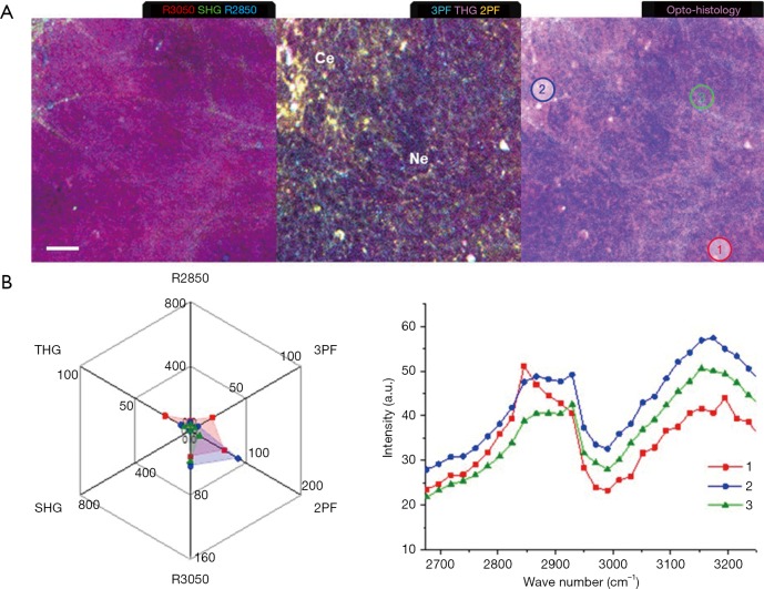
Figure 8.
Multiphoton imaging and molecular profiling for mammary tumor development in Week 9, demonstrating reduced cellular metabolism and collagen contents in a necrotic core. (A) Multimodal multiphoton images and the opto-histology. (B) Multiphoton molecular profiles of the selected areas in the opto-histology shown in (A). Morphological features—Ce, hypercellularity; Ne, necrosis. Scale bar: 25 µm.

