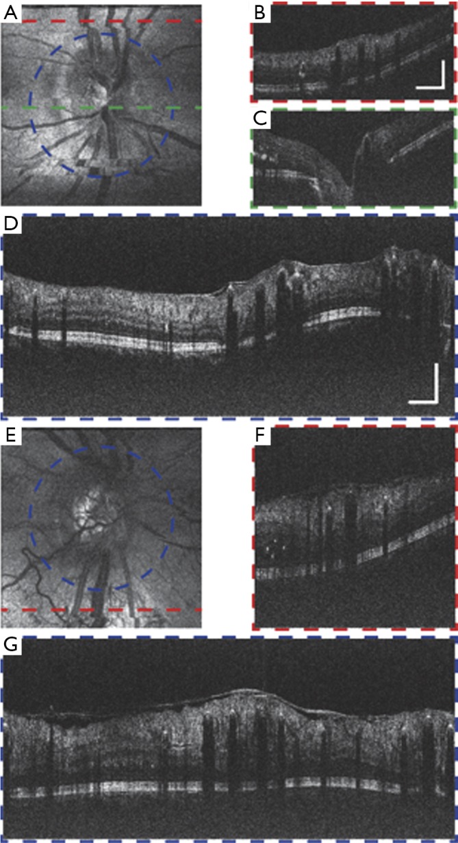
Figure 8.

Vis-OCT images of eyes with diabetic retinopathy (DR). (A,B,C,D) Images of a DR patient (57-year-old) (A) En face image of the region around ONH. (B) B-scan image along the red dashed-line in (A). Scale bar: 1 mm (lateral) by 0.2 mm (axial), applies to (A,B,C). (C) B-scan image along the green dashed-line in (A). (D) Circular B-scan image along the blue dashed-line in (A). Scale bar: 1 mm (lateral) by 0.2 mm (axial), applies to (D,E,F,G). (E,F,G) Images of another DR patient (38-year-old). (E) En face image of the region around ONH. (F) B-scan image along the red dashed-line in (E). (G) Circular B-scan image along the blue dashed-line in (E). No image averaging. Vis-OCT, visible-light optical coherence tomography; ONH, optic nerve head.
