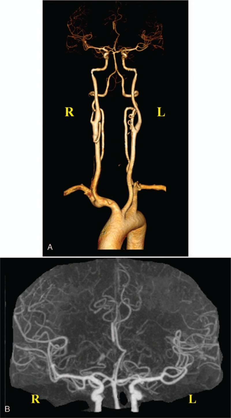
Figure 2.

Computed tomography angiography. (A) Three-dimensional imaging shows that the A2 segment of the right anterior cerebral artery is severely stenosed, along with basilar artery fenestration. The remaining vessels are naturally shaped with no stenosis or dilatation observed in the lumen. (B) Enlarged original image confirms severe stenosis of the A2 segment of the right anterior cerebral artery.
