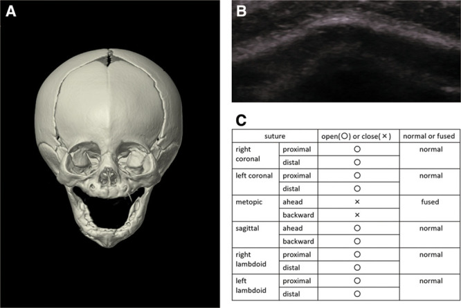
Fig. 7.

A 5-month-old girl with metopic craniosynostosis. A, Three-dimensional CT confirmed abnormal closure of the metopic suture. B, Ultrasound image showed disappearance of the hypoechogenic gap between the metopic bones. C, Ultrasound results are easy to visualize by filling out the table.
