
Figure 2.
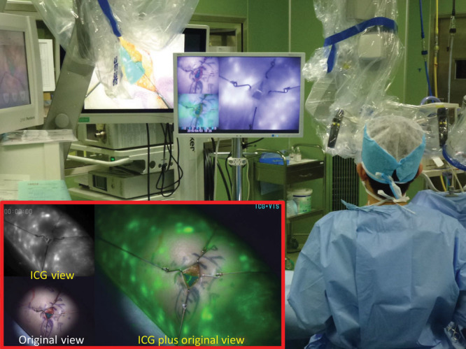
Detection of the lymphatic vessel in stardust patterns using real-time ICG videolymphography imaging. ICG fluorescence images are clearly visualized by an ICG videolymphography under the microscope illumination of the xenon light. The system monitors the operation field real-timely by 3 different simultaneous images: original view with visible light, which include no ICG fluorescence images, ICG view with near-infrared fluorescence light images, and ICG plus original view, which combines original view and ICG view with enhancing ICG fluorescence as green color in the image.