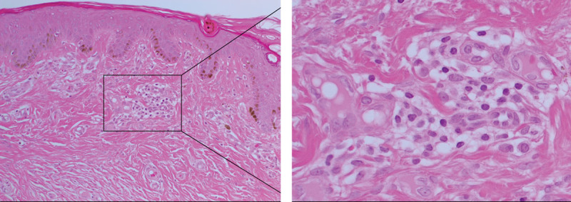
Fig. 1.

Hematoxylin & eosin staining of keloid tissue demonstrating the abundant collagen deposition within the dermis and the presence of the keloid-associated lymphoid tissues containing microvessels and inflammatory cells (inset), just beneath the epidermis. Original magnification: 200×.
