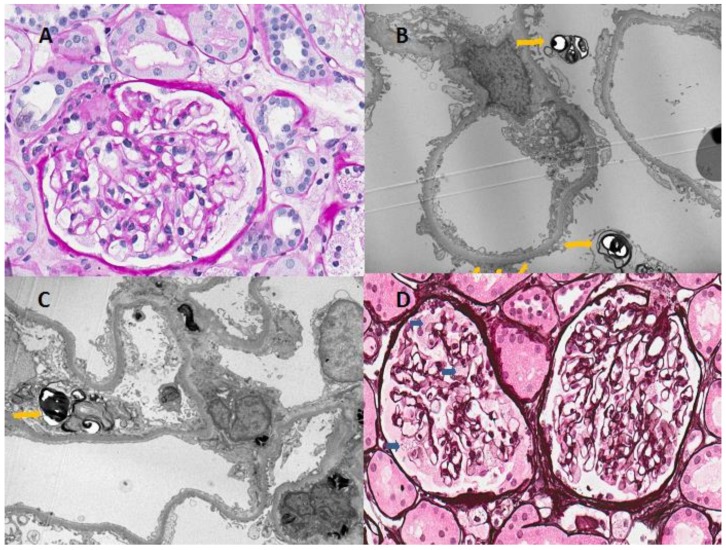
Figure 2.
Light and electron microscopic findings of renal biopsy from the patient. (A) Microscopic examination revealed minimal mesangial proliferative changes; (B,C) Typical findings of Fabry disease-multilamellated myelin figures (yellow arrowhead) are seen in the cytoplasm of podocytes on electron microscopy; (D) Light microscopy showed the vacuolization of podocytes (blue arrowhead), although the patient had normal renal function and nonsignificant proteinuria (Haemotoxylin and Eosin stain, ×400).

