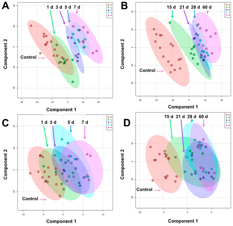
Figure 1.
PLS-DA plots comparing pre-exposure to (A) 1–7 d in urine, (B) 15–60 d in urine, (C) 1–7 d in serum, and (D) 15–60 d in serum after 4 Gy γ-ray TBI in NHPs. Overall, urine showed better separation among groups than serum. The highest separation for both biofluids occur within 15 d and higher overlap is observed from 21–60 d (graphs generated in MetaboAnalyst 4.0, pre-exposure samples [-8 and -3 d] were averaged for the control group).

