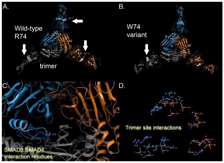
Figure 1.
SMAD3 molecular model for the full-length human sequence consisting of 425 amino acids and variant p.R74W. (A) Full-length model for the entire SMAD3 structure that shows trimer interaction with position 74 indicated by arrows. Each monomer of the trimer is indicated by a different color of ribbon (blue, orange, gray). (B) p.R74W variant for SMAD3. W74 is indicated by arrows. (C) Enlarged (zoomed into) the interaction region for SMAD3, where trimers are visible and also where SMAD4 can form heterodimers. Homodimerization and interaction residues are shown, colored by a SMAD3 monomer (blue, orange, gray). (D) "Ribbonless" view of the trimer interaction residues is shown. All protein residues shown in licorice rendering and using standard element coloring (O-red, N-blue, H-white, S-yellow), where carbons are shown in orange, blue or gray to indicate the monomer derived for trimer.

