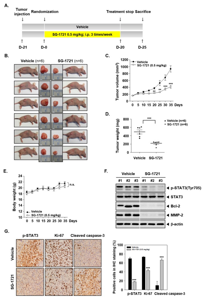
Figure 5.
SG-1721 inhibits tumor growth in vivo. (A) A schematic representation of experimental protocol described in “Materials and Methods”. (B) Necropsy photographs of mice bearing subcutaneously implanted triple-negative breast cancer (TNBC) cells. (C) The tumor diameters were measured at 5-day intervals, and the tumor volumes were calculated using the formula V = 4/3 πr3 (n = 6). (D) Tumor weight was measured during the experiment. (E) Body weight changes of mice were measured at indicated times. (F) Western blot of various proteins of interest was carried out in lysate from vehicle control and SG-1721 treated mice. (G) Immunohistochemical analysis of p-STAT3, proliferation marker Ki-67, and cleaved caspase-3 in the tumor tissues (left panel). The results shown are representative of the three independent experiments. Graphs represent positive cells in IHC staining (right panel).

