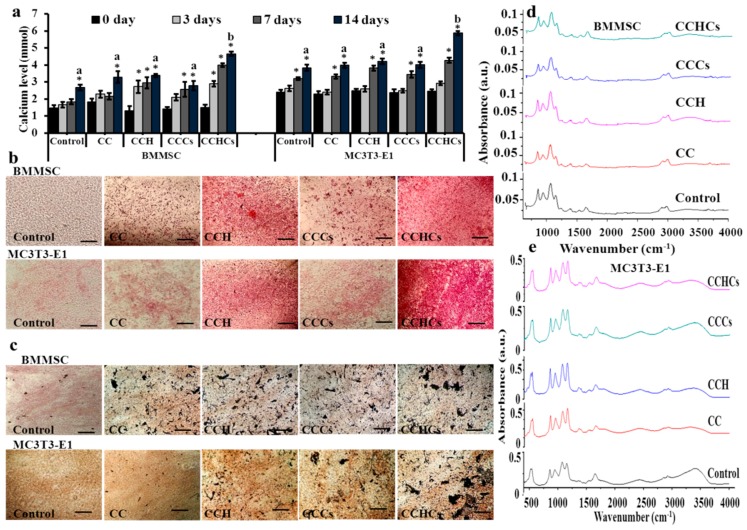
Figure 4.
Cellular calcium deposition of bone cells cultured on chitosan-collagen based bio-mimic 3D matrices for 14 days. The cellular mineral level (a), Alizarin red (b), and von Kossa (c) staining of bone cells. Scale bars: 40 μm. The presence of apatite was confirmed using FTIR spectra (d,e); control-cells grown on a regular tissue culture plate. BMMSC: bone marrow-derived mesenchymal stem cells; MC3T3-E1, pre-osteoblast. CC: chitosan-collagen 3D matrix, CCH: chitosan-collagen-hydroxyapatite 3D matrix, CCCs: chitosan-collagen-chondroitin sulfate 3D matrix, and CCHCs: chitosan-collagen-hydroxyapatite-chondroitin sulfate 3D matrix. The experiments were done three (a) or two (b–e) times with similar results. * p < 0.05 vs. zero days; different letters indicate statistical significance among 3D matrices for 14 days.

