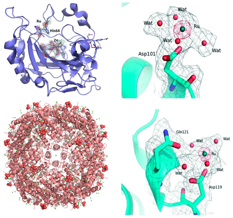
Figure 2.
Left: The adduct of NAMI-A with hen egg white lysozyme (HEWL). (top) Ru binding site close to Asp101. (bottom) Ru binding site close to Asp119. Reproduced from Ref. 59 by permission of The Royal Society of Chemistry. Top right: The adduct of NAMI-A with carbonic anhydrase (hCAII). Detail of the Ru center interactions with residues His 64. The oxygen atoms from water molecules are represented as red spheres. Reproduced from Ref. [60] with permission from Elsevier. Bottom right: Ribbon representation of the overall structure of the NAMI-A/HuHf (human ferritin) adduct. The side chain of His105 is shown as a ball and stick, while Ru and the water molecules completing the metal coordination sphere are shown as spheres. Reproduced from Ref. [61] by permission of The Royal Society of Chemistry.

