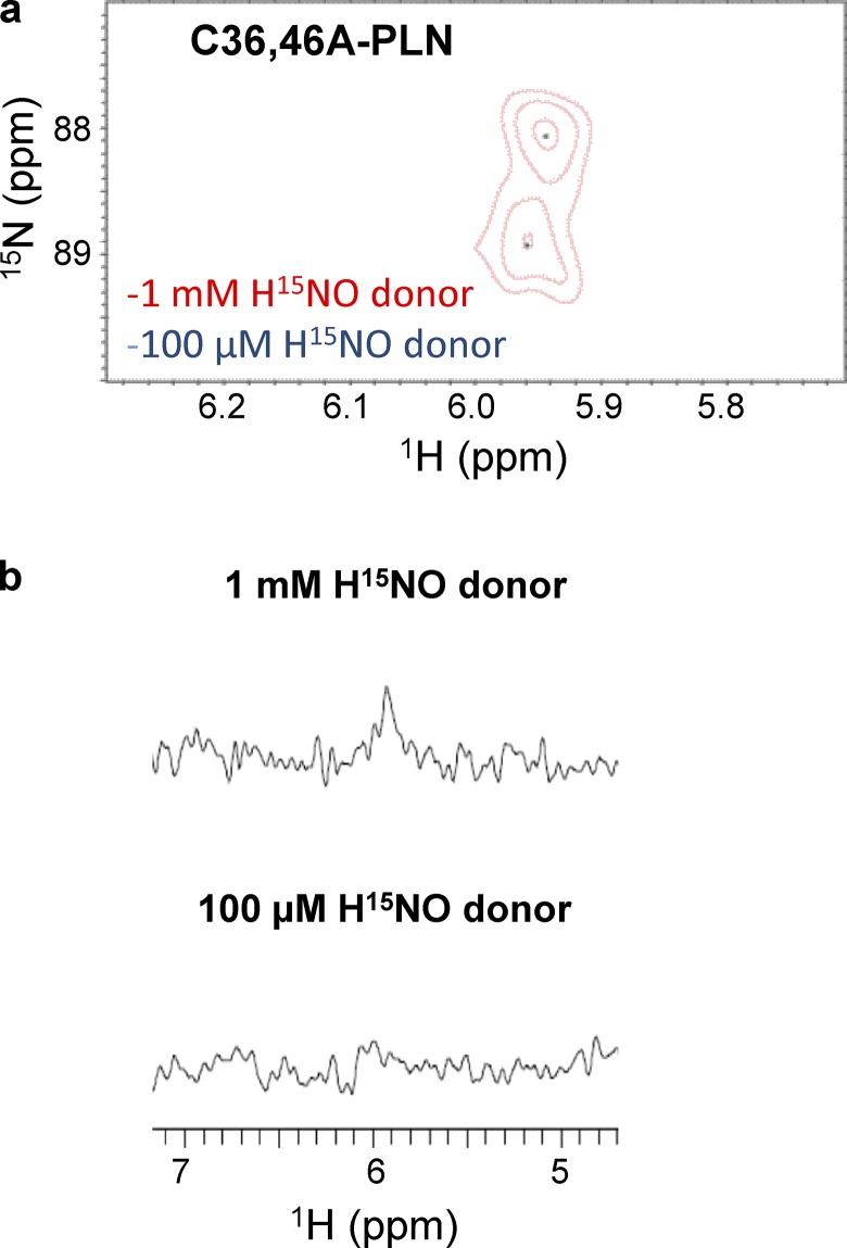
Figure 5.
Sulfinamide formation on C36,46A-PLN at different concentrations of HNO donor. (a and b) Comparison of the sulfinamide signals generated on C36,46A-PLN (60 µM, single Cys at position at 41) upon treatment with 1 mM (red) or 100 µM (blue) 15N-2-MSPA by 15N-edited 1H 2D-NMR (a) and 15N-edited 1H 1D-NMR analysis (b).

