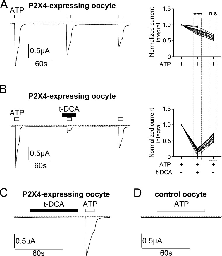
Figure 1.
Inhibition of ATP-activated P2X4-mediated currents by t-DCA. (A and B) Left panels: Representative whole-cell current traces recorded in human P2X4-expressing oocytes. ATP (10 µM) and t-DCA (250 µM) were present in the bath solution as indicated by open and filled bars, respectively. Zero current level is indicated by a dotted horizontal line. Right panels: Summary of results from similar experiments as shown in the left panels (A: n = 14, N = 1; B: n = 20, N = 2). Lines connect data points obtained in the same experiment. To quantify current responses, the integral of the inward current elicited by ATP application below the baseline was determined for each response. The current integral values of the second and third ATP-activated responses were normalized to the current integral of the first response in each individual recording (normalized current integral). ***, Second normalized responses in A and B are significantly different (P < 0.001). n.s., not significant. Third normalized responses in A and B are not significantly different; Student’s ratio t test. (C) Representative whole-cell current trace recorded in a human P2X4-expressing oocyte. ATP (10 µM) and t-DCA (250 µM) were present in the bath solution as indicated by open and filled bars, respectively. Zero current level is indicated by a dotted horizontal line (n = 25, N = 2). (D) Representative whole-cell current trace recorded in a control oocyte. ATP (100 µM) was present in the bath solution as indicated by open bar. Zero current level is indicated by a dotted horizontal line (n = 25, N = 2).

