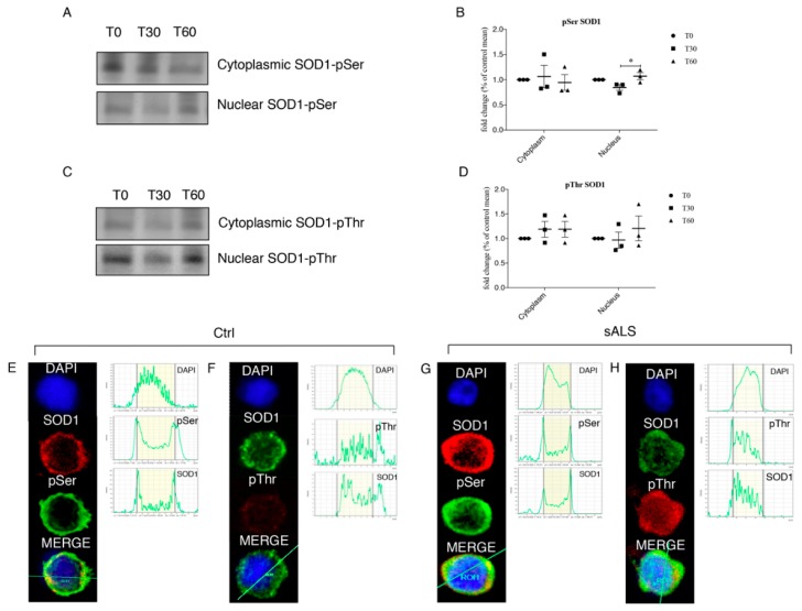
Figure 4.
(A,B) 1 mM H2O2 treatment determines significant phosphorylation at Ser residue at T60 in the nuclear compartment. SOD1 was immunoprecipitated and representative immunoblotting for pSer was reported for the nucleus. (C,D) 1 mM H2O2 treatment induces significant phosphorylation also at Thr residue at T60 in the nuclear compartment. SOD1 was immunoprecipitated and representative immunoblotting for pThr was reported for the nucleus. Data were analyzed by ANOVA (n = 3), followed by Newman-Keuls Multiple Comparison Test; * p < 0.05. PBMCs from healthy controls and from a subgroup of sALS patients were analyzed for pThr, pSer and SOD1 by immunofluorescence followed by confocal microscopy analysis. (E,F) In healthy controls both pThr and pSer were not observed in the nuclear fraction; (G,H) in PBMCs of sALS patients we observed a bright signal of SOD1 and pThr in the nuclear compartment, while SOD1 and pSer co-localization showed a slight signal. Nuclei were visualized using the fluorescent nuclear dye DAPI (blue).

