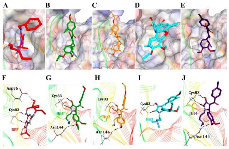
Figure 5.
Binding mode analyses. All the hit molecules preferentially lodged in the ATP-binding site of Cdk5 with almost similar molecular orientation (A–E). The REF, Hit1, Hit2, Hit3, and Hit4 are depicted as red, green, orange, cyan, and blue, respectively. The three dimensional (3D) molecular interaction pattern of the REF and all the hit molecules with Cdk5/p25 has been illustrated (F–J). Interacting residues are displayed as thin sticks and labeled. REF, Hit1, Hit2, Hit3, and Hit4 are depicted as thick stick representation and colored as red, green, orange, cyan, and blue, respectively. Hydrogen bonds have been shown as black-dashed lines.

