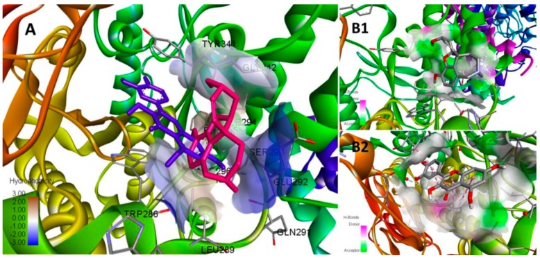
Figure 11.
Conformational analysis of galantamine and quercetin docked at the catalytic site of AChE; (A) Simulated best binding mode of galantamine (pink) and quercetin (blue) shares the same binding pocket with quercetin oriented more towards the hydrophobic domain. (B1) Galantamine stabilized its conformation mainly via a pattern of hydrogen bonding colored as green (donor) and pink (acceptor) sites at the pocket. (B2) The conformation of quercetin is stabilized by the pattern of hydrophobic interactions (gray site) in addition to H-bonding at the pocket.

