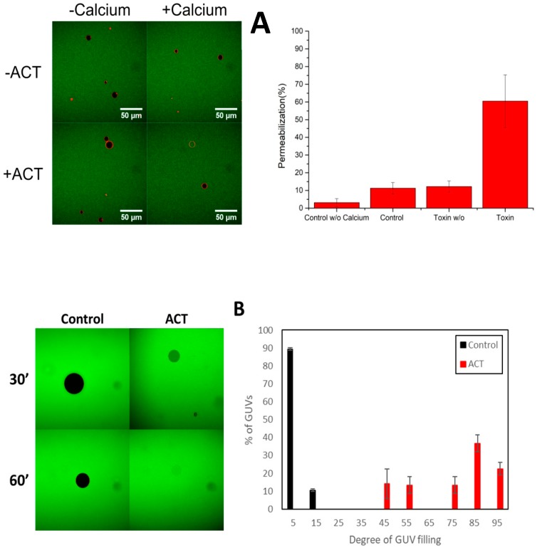
Figure 1.
Calcium-dependent-permeabilization of dioleylphosphatidylcholine Giant Unilamellar Vesicles by ACT DOPC GUVs by ACT toxin. (A) Representative images of GUVs (black, if empty) in a solution of Alexa-Fluor488 (green) incubated in the absence or presence of 200 nM ACT and buffer without or with 10 mM CaCl2 (left panel). Images were taken 30 min after mixing the components. The internalization of Alexa-Fluor-488 to the lumen of GUVs (green) corresponds to permeabilized vesicles. GUV composition was DOPC. In the right panel the total percentage of permeabilization after 30′ for each condition (± ACT ± CaCl2) is depicted. In this case the threshold filling value to discriminate between permeabilized and non-permeabilized vesicles has been the 40%. (B) Distribution of the degree of filling of individual GUVs to Alexa-Fluor-488 (right panel), after treatment with 200 nM ACT for 30 min in buffer with 10 mM CaCl2. The degree of filling was calculated for each individual vesicle, from confocal images as the one shown in the left pane. In each of three experiments, a minimum of 250 vesicles were analyzed per condition. Error bars represent S.D.

