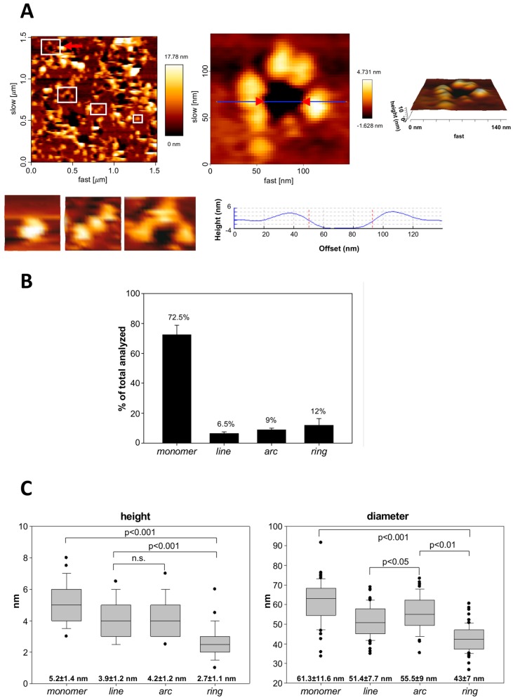
Figure 5.
Analysis by atomic force microscopy of the assemblies formed by ACT in supported lipid membranes. (A) AFM image (left upper panel) of a supported lipid bilayer (SLB) prepared from ACT-containing proteoliposomes (POPC liposomes reconstituted with ACT). The red arrowhead points to a membrane pore that has been selected for a more detailed topographic analysis; in the image there are more pores, heterogeneous in size and shape; the edges of the pores present protrusions corresponding to ACT clusters. Below this image, other ACT assemblies are shown, such as a monomer, a line and an arc, which have been selected from other AFM images. On the right-hand part of panel A, a 3D AFM topography of the pointed ACT ring is shown in a greater detail. The image reveals a circular dark hole that spans the lipid membrane (below). ACT molecules around the pore rim (yellow-white) protrude 3.97 ± 1.02 nm above the membrane plane, as confirmed by the height profile shown below the image (corresponding to the blue line in the middle of the 2D image. (B) Quantitative analysis of the structures found for ACT on SLBs. Data show the percentage of each type of structure (monomer, line, arc or ring) in all the measurements. (C) Determination of the diameter and height (in nm) of the monomeric particle in each of the different ACT assemblies (monomer, line, arc or closed ring). Mean values are depicted as box-and-whisker plots (the ends of the whiskers represent standard deviations). A total of 973 particles were measured for these determinations.

