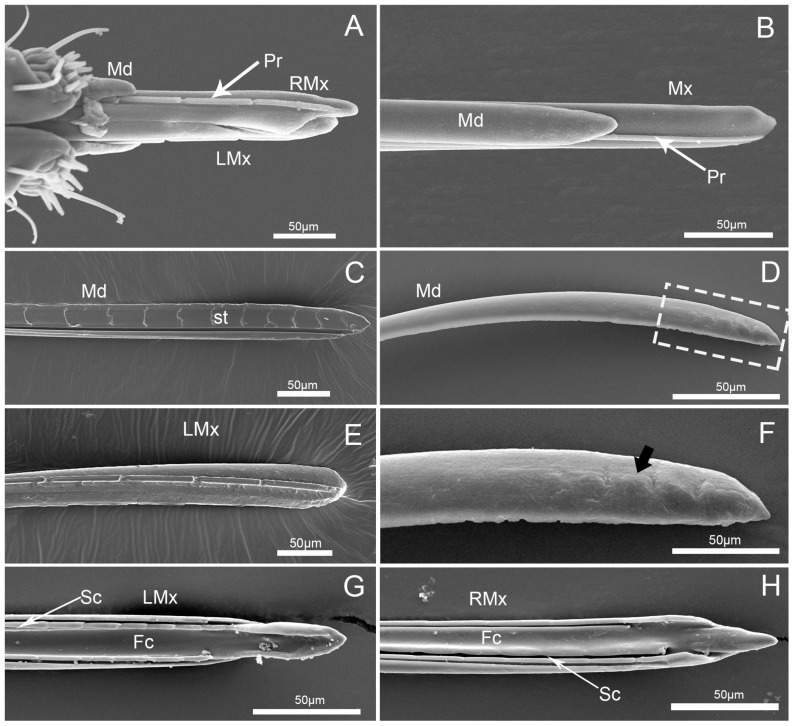
Figure 8.
SEM images of stylet of C.nigrescens. (A) Enlarged anterior view of apex of stylet fascicle; (B) enlarged view of stylet fascicle; (C) interior view of mandibular stylets (Md); (D) external view of mandibular stylets (Md); (E) external view of left maxillary stylet (LMx); (F) enlarged view of the box in (D), showing notches (arrow); (G) interior view of left maxillary stylet (LMx); and (H) interior view of right maxillary stylet (RMx); Fc, food canal; Pr, process; Sc, salivary canal; Sr, serrate ridges; and st, squamous texture.

