Fig. 1.
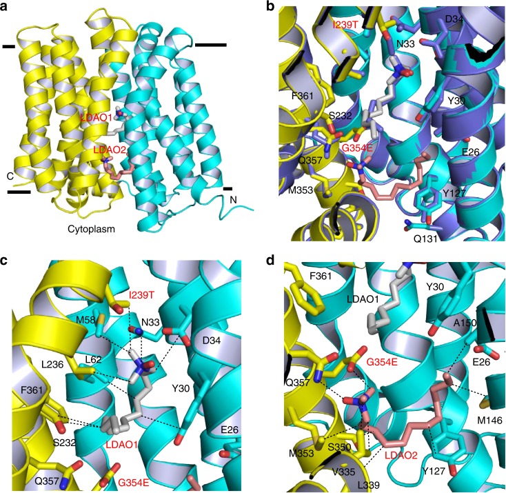
Structure of the LDAO-bound I239T/G354E. a Structure of I239T/G354E as viewed from the membrane. I239T/G354E is drawn as a ribbon diagram, with the N (9–205) and C (206–400) domains colored cyan and yellow, respectively. The bound LDAO molecules are shown as stick models and are colored grey and light pink, respectively. b Overlay of the LDAO-bound structures of Q131R (light blue, PDB 4ZP2) and I239T/G354E, with the LDAO-binding amino acids and LDAO drawn as stick models. The Q131R-bound LDAO is colored light blue. Q131R was modeled as alanine in 4ZP2; in I239T/G354E, Q131 makes no contact with the bound LDAO molecules. c Close-up view of the LDAO1-binding site, LDAO1 and the relevant amino acids are shown in stick models and close-range interactions are highlighted as dashed lines. d Structure of the LDAO2-binding site, LDAO2 and the relevant amino acids are drawn as sticks, dashed lines indicate the interactions between LDAO2 and I239T/G354E

