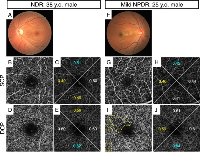
Figure 1.
Representative cases of an eye without DR (NDR, 38 y.o. male, HbA1c 6.5%, DM duration 2 years; (A–E) and an eye with DR (mild NPDR, 25 y.o. male, HbA1c 7.7%, DM duration 1 year; (F–J). This NPDR case showed local capillary dropout and higher orientation bias compared with the NDR case although whole flow density (FD) in NPDR was not decreased as compared with that of NDR. (A,F): color fundus photograph, (B,C,G,H): 2.6 × 2.6 mm SCP images, (D,E,I,J): 2.6 × 2.6 mm DCP images. Numbers indicate FD of each area in (C,E,H,J). The FD of the whole area in (C,E,H,J) was 0.50, 0.60, 0.43, and 0.61, respectively. Yellow dotted circles indicate capillary dropout (I).

