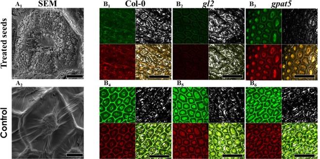
Figure 4.
Low temperature plasma affects A. thaliana seeds surface. (A) Col-0 seed columella with or without plasma treatment observed with a scanning electron microscopy. Scale bar: 10 µm. (B) Confocal observation of seed from the three different A. thaliana genotypes with or without plasma treatment using lens 10X zoom x9. The seeds are previously stained with auramine-O. After excitation at 488 nm, seed surface was visualized by reflection at 484–494 nm (gray); by fluorescence at 505–560 nm (green), by fluorescence at 572–642 nm (red). The right bottom panel corresponds to the merged of the 3 detection. Scale bar: 100 µm.

