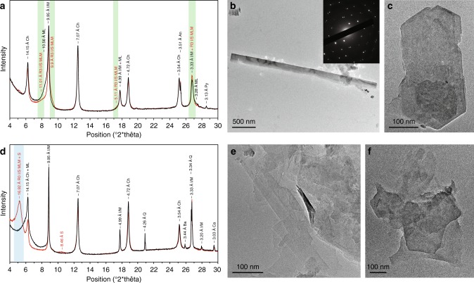
Fig. 5.
Mineralogical composition of the <2 µm clay-size fractions. X-ray diffraction profiles of oriented preparations after air-drying (black lines) and glycolation (red lines) and their transmission-electron images are given. a Microbial mat specimen. b Large, well-crystallized lath from microbial mat laminae. The inset shows selected area electron diffraction (SAED) pattern. hk0 pattern shows a hexagonal structure typical of phyllosilicates and coherently stacked crystals. c Hexagonal habit of illite from a mat sample. d Associated sandstone sediment. e Lathlike and poorly crystallized particles from the host sandstone. f Tiny hexagonal-shaped particles from the host sediment. [Green areas correspond to long-range ordered illite–smectite mixed-layer minerals (R3 I/S MLM); Blue areas represent randomly ordered illite–smectite mixed-layer minerals (R0 I/S MLM); smectite (S); chlorite (Ch); mixed-layer (ML); illite/mica (I/M); anatase (An); quartz (Q); pyrite (Py); barite (Ba); calcite (Ca)]

