
Figure 2.
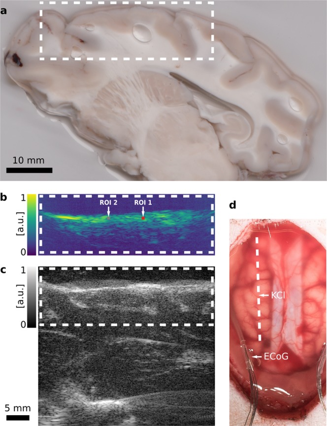
Hybrid photoacoustic ultrasonic (PAUS) imaging of a porcine brain. The dashed white line and boxes show corresponding sections of the swine cortex. (a) Photograph of a sagittal surgical slice segmented from the extracted brain, 1 cm from the midline. The shown segment is manually registered to (b) a representative photoacoustic (PA) image with two regions of interest (ROI), and (c) the corresponding ultrasound (US) B-Mode image. Both the US and the PA image are shown with in arbitrary units with logarithmic compression. (d) Photograph of the exposed cortex after the craniotomy and dura mater retraction with the dashed line marking the PAUS imaging plane. The electrocorticography (ECoG) electrodes are positioned in the lateral margins. The location of the KCl stimulation is denoted as well.