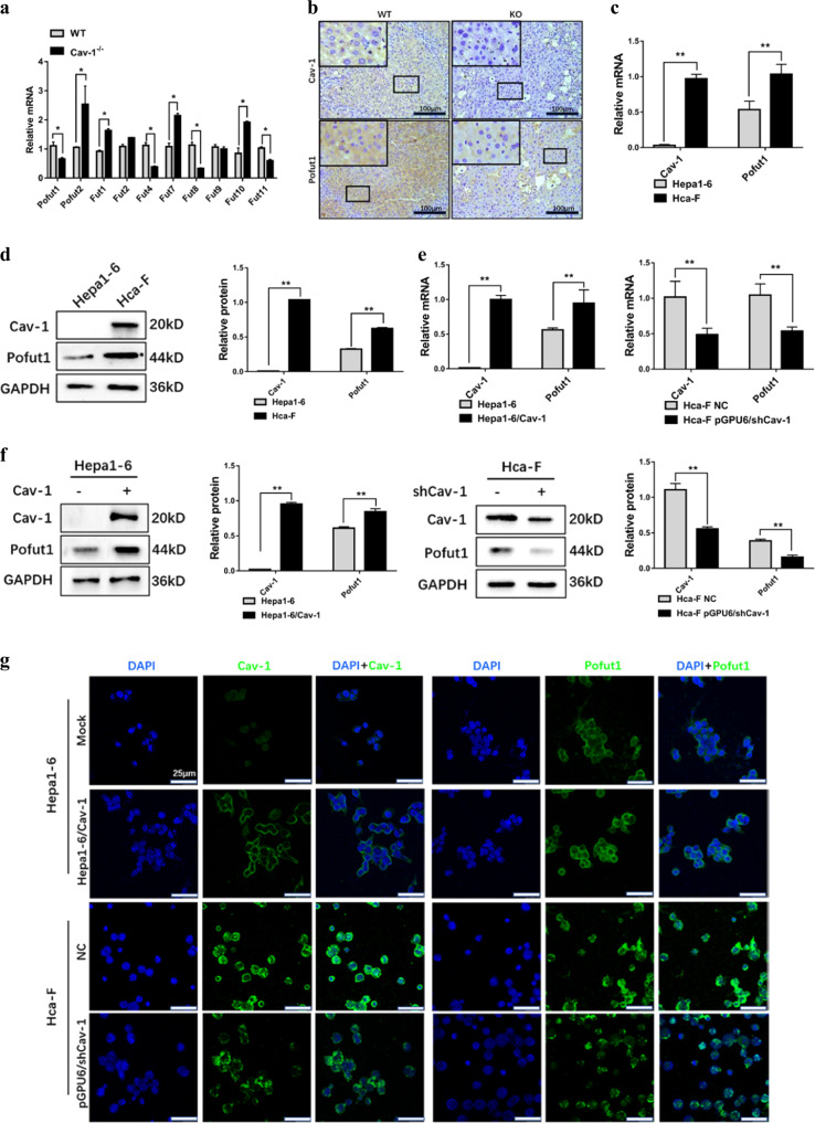
Fig. 1. Pofut1 expression is positively associated with Caveolin-1 (Cav-1).
a Analysis of the mRNA levels of different fucosyltransferases in wild-type C57BL/6J mice and Cav-1−/− C57BL/6J mice. b Expression levels of Pofut1 were examined by immunohistochemical staining. The blue staining represents the nucleus, and brown staining represents Cav-1 and Pofut1 (the secondary antibody was labeled with horseradish peroxidase, which produces a brown color with the substrate DAB). Representative sections of liver tissues from wild-type and Cav-1−/− C57BL/6 mice. Original magnification, ×100. c Endogenous Cav-1 and Pofut1 mRNA levels in Hepa1-6 and Hca-F cell lines were determined by quantitative real-time PCR (qRT-PCR). GAPDH served as a loading control. d Endogenous Cav-1 and Pofut1 protein levels in Hepa1-6 and Hca-F cell lines were determined by western blotting. GAPDH served as a loading control. e qRT-PCR assay was used to detect the mRNA expression level of Pofut1 after overexpression of Cav-1 in Hepa1-6 cells and after knockdown of Cav-1 in Hca-F cells. GAPDH served as a loading control. f Western blot assay was used to detect the protein expression level of Pofut1 after overexpression of Cav-1 in Hepa1-6 cells and after knockdown of Cav-1 in Hca-F cells. GAPDH served as a loading control. g Immunofluorescence staining was performed to further detect the protein expression of Pofut1 after overexpression of Cav-1 in Hepa1-6 cells and after knockdown of Cav-1 in Hca-F cells. Nuclei were counterstained with DAPI (×40). Statistical comparison using t test: *P < 0.05, **P < 0.01

