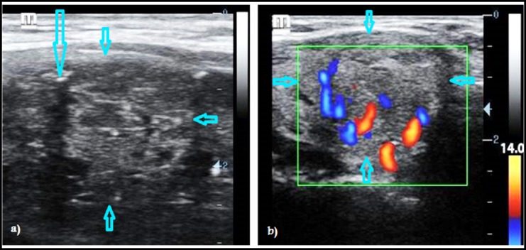
Fig.2.

Ultrasound images of thyroid nodules in two different patients. B-mode ultrasonography showing a well-defined round isoechoic nodule with smooth borders but a fleck of micro-calcification (long arrow) inside it give suspicious of malignancy (a). Power Doppler image (PDI) of another patient showing a well-defined round slightly hypoechoic nodule with smooth borders and central vascularity that give suspicious of malignancy (b).
