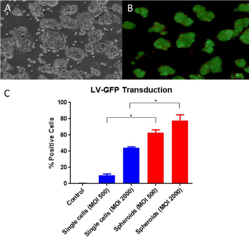
Fig. 2.

Morphology and viability of spheroids, and differences in transduction efficiency between single-cell and spheroid hepatocytes. (A) Light microscopy image of spheroids. (B) Inverted fluorescence microscopy image of spheroid hepatocytes, using the Fluoroquench viability stain before transplantation in pig 897; live cells (green) and necrotic cells (orange). (C) Ex vivo transduction of primary pig single-cell hepatocytes and hepatocyte spheroids with LV-GFP at MOls of 500 and 2000 LPs per cell and quantification of GFP-positive cells detected by flow cytometry 96 hours after transduction. Data are means ± SD ( n=3 replicates for single cells, 2 replicates for spheroids). *P < .01.
