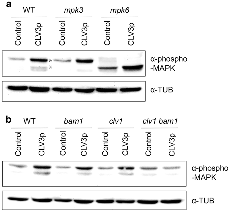
Fig. 2.
MPK3 and MPK6 are activated by treatment with CLV3p signals. a Immunoblot assays using the shoot apex tissues harvested from WT Col-0, mpk3 and mpk6 mutant seedlings treated without (Control) or with 10 μM CLV3p for 10 min. Asterisks, endogenous MPK3 and MPK6. b Immunoblot assays using the shoot apex tissues harvested from WT Col-0, bam1, clv1 and clv1 bam1 mutant seedlings treated without (Control) or with 10 μM CLV3p for 10 min. Activated MAPKs were detected by an α-phospho-MAPK antibody and loading amounts of proteins were detected by an α-TUB antibody. These experiments were repeated at least three times with similar results

