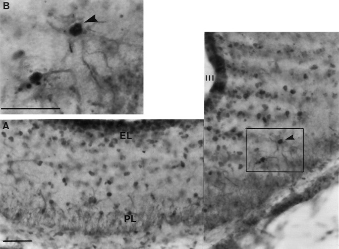
Fig. 6.
FLI-labeled glia are widespread throughout the ME after photostimulation. A, The composite photomicrograph was taken at the level of the ME of a quail killed after 18 hr of light. B, A high power example is shown in theinset micrograph; an example of an activated glial cell (as demonstrated by FLI) within the ME is indicated by thearrowhead. The darker-labeled FLI nuclei can be located within the centers of the lighter cytoplasmic-stained glial cells.III, Third ventricle; EL, ependymal layer of the ME; PL, palisade layer of the ME. Scale bars, 25 μm.

