
Fig. 1.
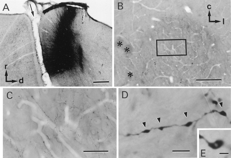
Identification of corticorubral fibers.A, Injection site of biocytin. A sagittal section of the sensorimotor cortex is shown. The arrow indicates the cruciate sulcus. r, Rostral; d, dorsal.B, Low-magnification photomicrograph of a horizontal section of the RN. Asterisks indicate the oculomotor nerve. c, Caudal; l, lateral.C, High-magnification photomicrograph of the area of the RN outlined by the rectangle in B. Many biocytin-labeled fibers can be seen. D,E, Higher-magnification photomicrographs of biocytin-labeled fibers. Axonal swellings are seen along the fibers (arrowheads). Some of the axonal swellings were fenestrated, as shown in E. Scale bars:A, 500 μm; B, 200 μm;C, 50 μm; D, 4 μm; E, 2 μm.