
Fig. 7.
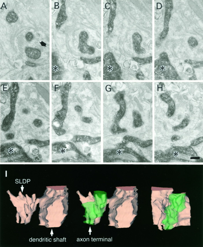
SLDPs of an HRP-labeled RS cell invaginating into an axon terminal. Asterisks show the dendritic shaft from which SLDPs emanate. The black arrow shows the synaptic terminal. A–H, Electron micrographs of selected serial sections. I, Three-dimensional reconstruction of the presynaptic axon terminal and the SLDP. See legend of Figure 3 for detail. Scale bar, 0.25 μm.