
Fig. 9.
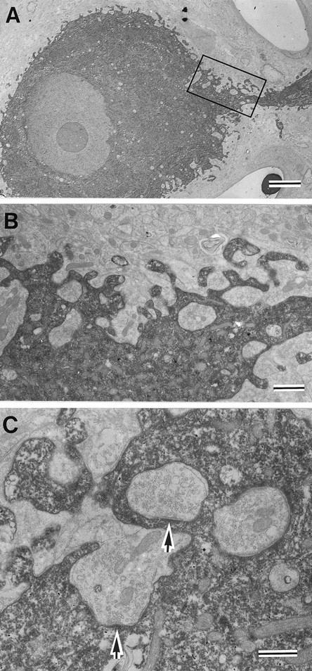
Synapses on the somatic membrane of RS cells.A, Electron micrograph of an HRP-filled RS cell.B, Higher magnification of the area outlined by therectangle in A. Note the presence of inclusions of synaptic endings in the soma. C, Synapses formed by terminals included in the soma (arrows). Similar inclusions were observed in nonstained soma, indicating that these are not artifacts of the HRP injection (not shown). Scale bars:A, 5 μm; B, 1 μm; C, 0.5 μm.