
Fig. 2.
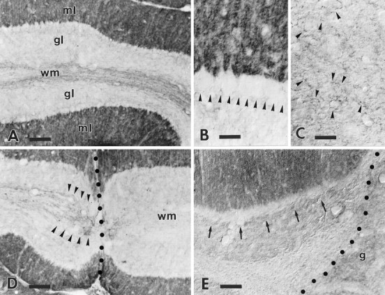
B-50/GAP-43 expression in intact and injured wild-type cerebella. The survey micrograph A shows the anti-B-50/GAP-43 labeling pattern in the wild-type cerebellum. The molecular layer (ml) shows an intense punctate labeling, whereas the granular layer (gl) is almost completely unlabeled. The row of virtually immunonegative Purkinje cell somata at the granular–molecular layer interface is highlighted by the contrast with the strongly labeled molecular layer. Note the presence of several dimly labeled axons running along the axial white matter (wm) of this folium. The higher magnification in B shows the absence of staining in Purkinje cell perikarya (arrowheads). Labeling in the deep nuclei (C) is restricted to sparse varicose branches (some are indicated by arrowheads) displaying a faint immunoreactivity. The labeling pattern in the cortex is substantially unaltered in the transected folia (D): Purkinje cells remain immunonegative. Note that all immunoreactive axons (arrowheads) stop abruptly at the lesion site (dotted line), and no labeled profiles remain in the white matter (wm) on the other side of the injury. A similar picture is observed when the transected folia face a transplant (E), a cerebellar graft (g) in this case: arrows point to the immunonegative Purkinje cell somata (dotted lineindicates the graft–host border). Survival times: 21 d (D), 60 d (E). Scale bars: A, D, 90 μm; B, C, 30 μm;E, 60 μm.