
Fig. 6.
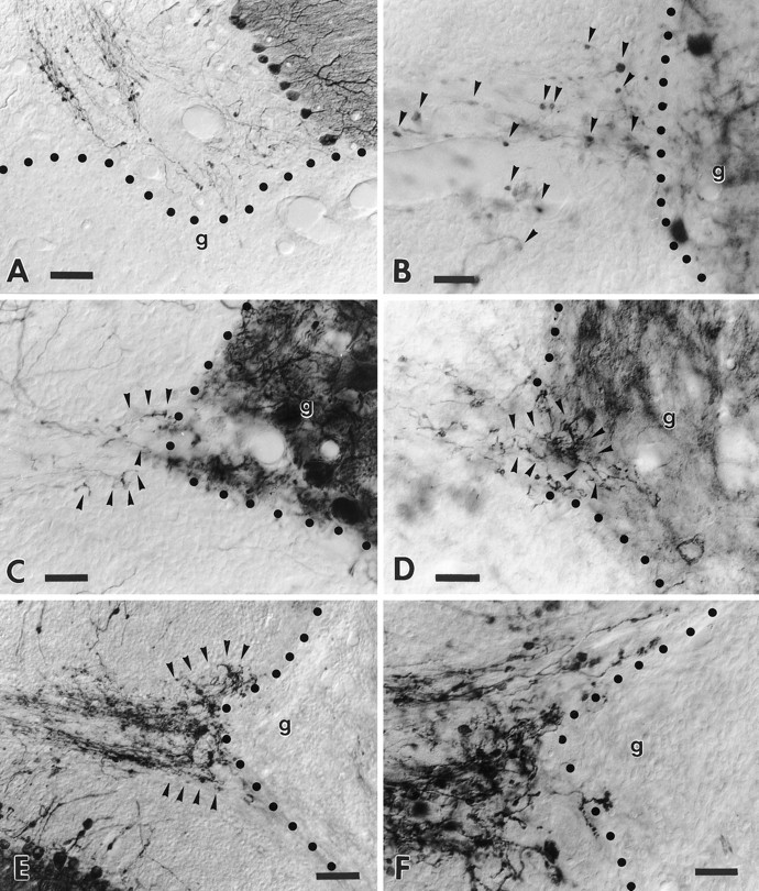
Transgenic Purkinje cell axons are unable to regenerate into growth-permissive transplants. A shows a transected folium from a transgenic cerebellum facing a large embryonic neocortical transplant. Note the anti-L7-immunolabeled axons that end at the graft–host interface. The high-magnification picture (B) shows anti-calbindin-immunolabeled wild-type Purkinje cell axons that terminate with round-shaped terminal clubs (arrowheads) close to the edge of an embryonic cerebellar transplant, highlighted by the presence of grafted Purkinje cells. By contrast, the micrograph C shows a similar situation in a transgenic animal also stained by anti-calbindin antibodies: several thin sprouts (arrowheads) emanate from the transected host axons. Anti-B-50/GAP-43 immunolabeling of the adjacent section (D) shows several sprouts (arrowheads) that elongate for a short distance into the transplant. Note, however, that these sprouts do not show the morphology of terminal varicose branches. The micrographE displays a prominent plexus (arrowheads) of anti-B-50/GAP-43-immunolabeled sprouts abutting a Schwann cell graft. The higher-magnification pictureF shows the typical morphology of the newly formed sprouts that abruptly stop at the graft–host border. In all picturesg indicates the graft, whereas the dotted line highlights the graft–host interface. Survival times: 30 d (A); 60 d (B–F). Scale bars: A, E, 60 μm; B, C, D, F, 30 μm.