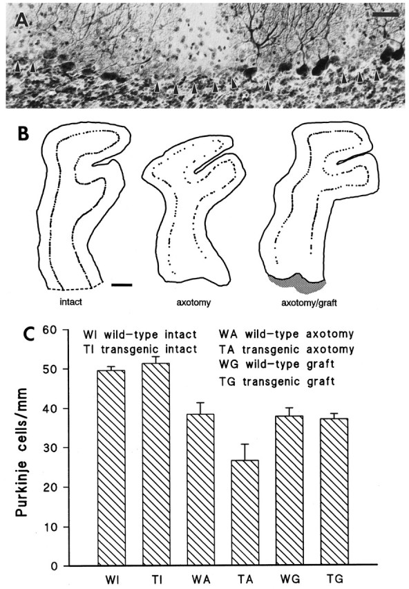
Fig. 7.

Purkinje cell loss in the injured wild-type and transgenic cerebella. A transected transgenic cerebellar lobule, immunolabeled by anti-L7 antibodies and counterstained by thionine, is displayed in A. Arrowheads point to areas where Purkinje cells have degenerated. Note that all surviving neurons are immunolabeled and no immunonegative thionine-stained Purkinje cell perikarya remain. B shows three representative camera lucida reconstructions of lobuli IV–V from transgenic mouse cerebella. Two months after axotomy (center) the number of Purkinje cells is considerably decreased compared with control (left). By contrast, when a graft (shaded area on the right: axotomy/graft) is placed in the lesion track, the injured cells are partially rescued. Quantitative estimations of the number of Purkinje cells/millimeter of Purkinje cell layer are shown in the histogram C. Intact wild-type and transgenic mice have similar values, indicating that no spontaneous cell loss occurs in transgenic animals. By contrast, after axotomy the number of Purkinje cells/millimeter is reduced in both animal sets, although a statistically more severe cell loss is observed in transgenic animals. Finally, when a graft is placed into the lesion site, both wild-type and transgenic mice show similar values that are not statistically different from that obtained from axotomized wild-type cerebella, indicating that axotomy-induced Purkinje cell loss in transgenic mice can be prevented by graft-derived trophic support. Scale bars: A, 40 μm; B, 200 μm.
