
Fig. 1.
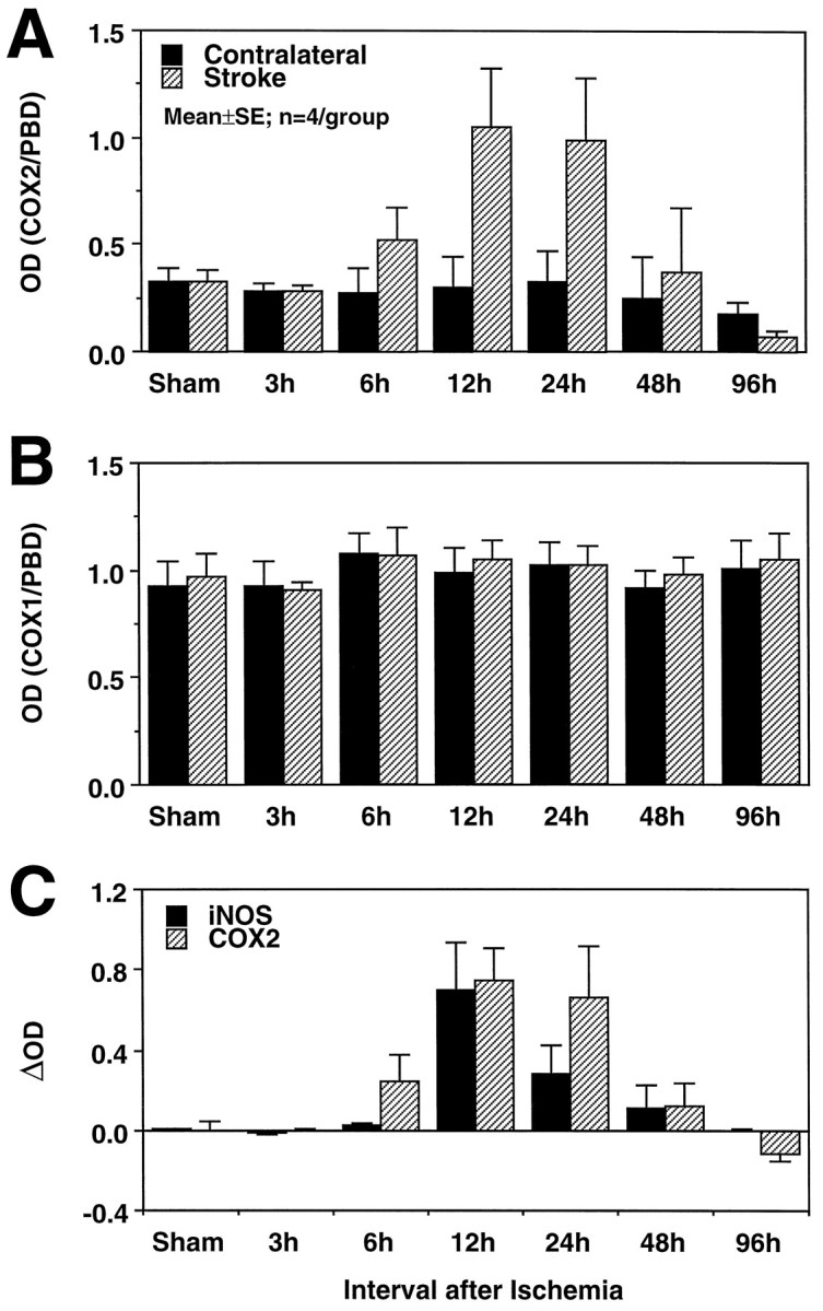
A, Effect of transient focal ischemia on COX-2 mRNA expression detected by RT-PCR. PCR products were run on a gel, and the optical density of the bands was measured by image analysis. The density of the COX-2 band was divided by the density of the band of a ubiquitous gene, PBD, used as a normalization factor. Each time point represents the average of four rats. The COX-2 signal increases at 6 hr, reaches a peak at 12–24 hr, and returns to baseline at 48–96 hr. No changes in COX-2 expression are seen in the brain contralateral to the stroke. B, In contrast to COX-2, COX-1 mRNA does not increase after cerebral ischemia on either side of the brain. The density of the COX-1 band was normalized by the PBD band as described in A. The fact that the mRNA for COX-1, an enzyme closely related to COX-2, was not increased attests to the selectivity of the COX-2 upregulation and to the selectivity of the RT-PCR technique used in the present study. C, Comparison of the time course of COX-2 and iNOS expression after transient cerebral ischemia. Data are presented as δ, obtained by subtracting the density of the COX-2 or iNOS bands in the contralateral nonischemic side from that of the stroke side. The time course of COX-2 and iNOS expression in the postischemic period is similar.