
Fig. 1.
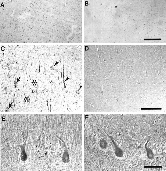
Nitrotyrosine immunoreactivity is prominent in the cytoplasm and nuclei of hippocampal neurons in cases of Alzheimer’s disease (A, C), whereas it is absent from control cases (B, D). Immunoreactivity was often but not always more intense in neurons containing NFTs (arrows) than in those lacking NFTs, which were also intensely stained (arrowheads). In contrast, amyloid-β deposits (✻) and surrounding dystrophic neurites of senile plaques as well as extracellular-NFTs (unmarked) were unstained. The location of NFTs and amyloid-β deposits was determined by Congo red counterstaining. In contrast, in the cerebellum, nitrotyrosine immunoreactivity was present at the same level in cases of Alzheimer’s disease (E) as it was in controls (F). Scale bars: A, B, 500 μm; C, D, 100 μm; E, F, 50 μm.