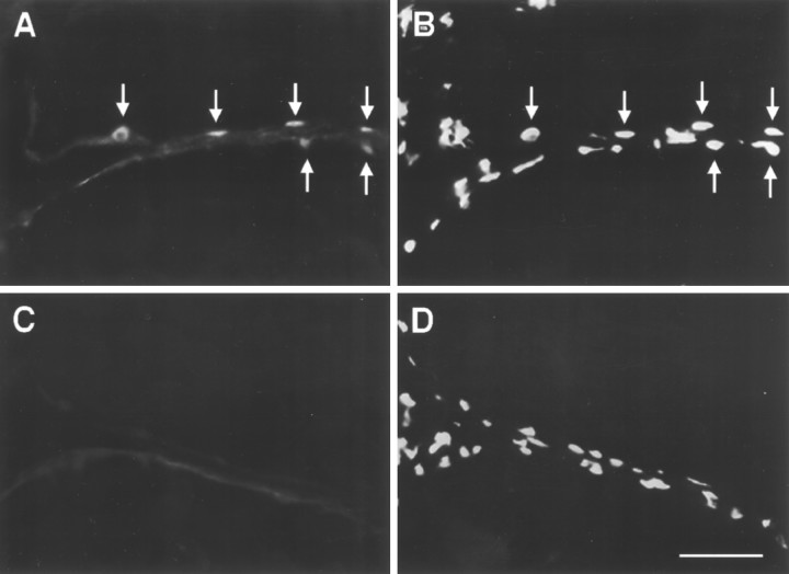
Fig. 4.
Immunocytochemical localization of ERs in the electric organ of Sternopygus. The fish was implanted with E2, and the tail was cut in cross section. Each photograph shows the intersection of two adjacent electrocytes.A, Section stained with antibody to ER (H222).Arrows indicate positively stained nuclei located at the periphery of adjacent cells. B, Same section as inA stained with propidium iodide to show all nuclei present. Arrows point to nuclei stained inA. Note that not all nuclei stain positively with H222.C, Control section incubated without primary antibody to show nonspecific staining of the secondary antibody. D, Same section as in C stained with propidium iodide. Scale bar, 50 μm.

