
Fig. 4.
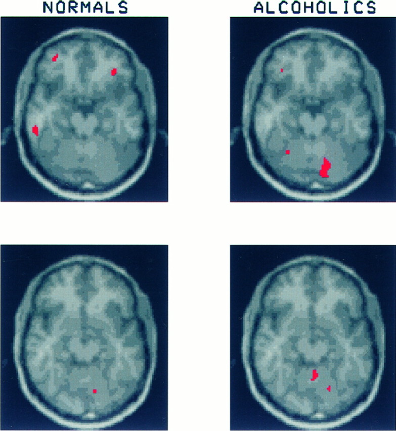
These slices through the superior aspects of the orbital cortex and cerebellum show three regions of significant activation in the healthy volunteers: one in the inferior temporal lobe on the left and bilateral regions in lateral orbital gyrus. Because these regions were relatively small and did not appear on contiguous slices, their mean CMRglc was not calculated. The area of significant activation seen in the right cerebellar hemisphere of the alcoholics was included in the calculation of mean CMRglc for the right cerebellar region described in Figure 3.