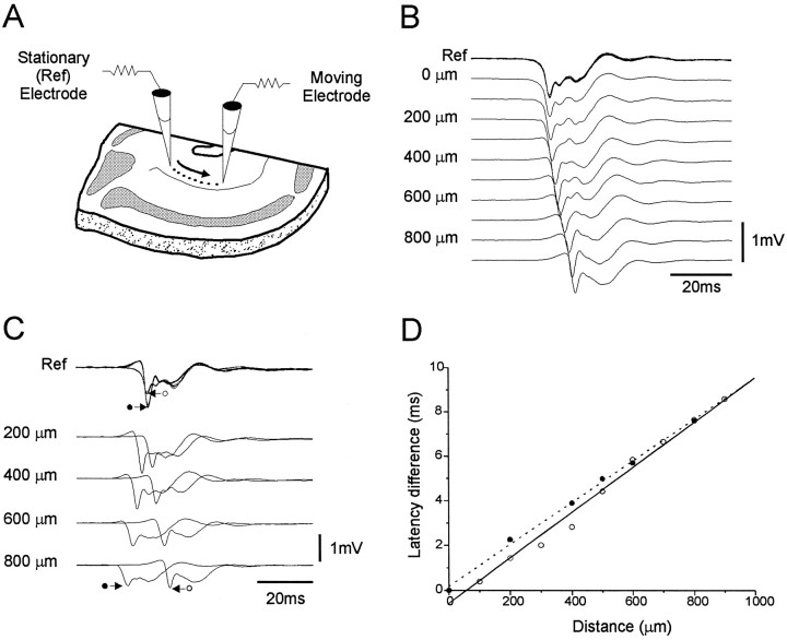
Fig. 3.
Horizontal propagation of epileptiform events in the entorhinal slice. A, Representation of the experimental protocol. A reference electrode was placed in the medial aspect of layers V–VI while a moving electrode was advanced in 100 μm increments along the medial-lateral axis of layers V–VI.B, Horizontal profile of the first 60 msec of an SDE. The traces at the top represent the superimposition of the potentials recorded at the reference electrode site for each of the horizontal levels shown below. As the moving electrode was moved further in a lateral direction the latency difference increased, indicating a medial to lateral propagation of the event. C, Combined horizontal profile for two distinct SDEs as distinguished by their different waveforms as recorded at the level of the stationary reference electrode (A). The events alternated in their occurrence and appeared to propagate in opposite directions. One event is the same as in B (○) and propagated in the medial-to-lateral direction as indicated by the increasing latency from the reference at increasing recording distances. The second (•) propagated in the lateral-to-medial direction as suggested by the opposite temporal relation. D, A regression plot of the relationship between the latency differences between the activity at the stationary and the moving electrode for both SDEs shown inC. Both events appear to travel at similar speeds across the horizontal aspect of the slice. The average propagation speed of the event shown in B (○) was 111 mm/sec, whereas the second event shown in C (•) propagated at 100 mm/sec.

