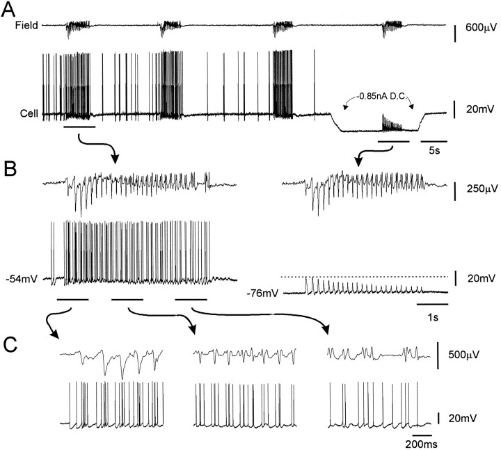
Fig. 6.
Activation of an electrophysiologically identified EC layer II SC during rhythmic epileptiform events evoked by CCh (20 μm). The field recording location was in layer II, 150 μm lateral to the intracellular electrode. A, Long trace showing the cell becoming strongly activated during epileptiform events. This cell was depolarized by 6 mV in response to CCh before the initiation of field events. During the last field event shown, DC hyperpolarization unveiled large-amplitude depolarizing synaptic potentials synchronized with the negative field spikes.B, Fast sweep speed trace of the first and last epileptiform events shown in A. C, Higher sweep speed traces for the indicated periods of the epileptiform event shown in B.

